In-House Methodologies
Establishing and validating cellular model systems
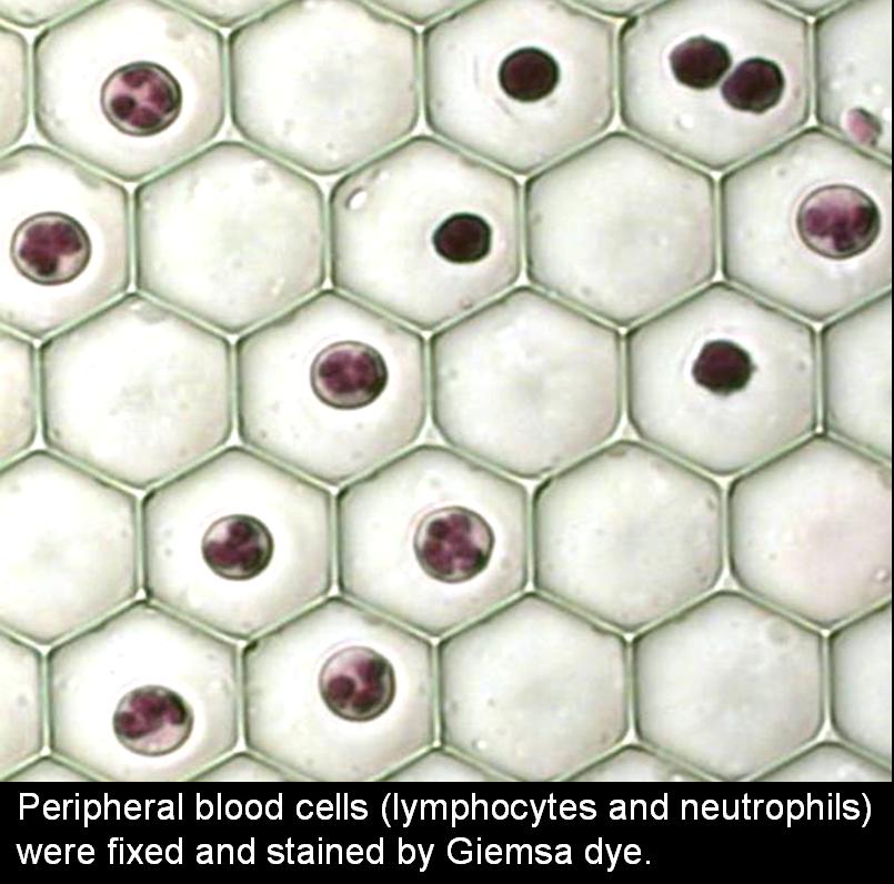
Various types of non-adherent cells (in suspension) of various sizes (peripheral lymphocytes, the Jurkat T cell line, Molt 4 T cell line, U937 and THP monocyte cell lines), as well as several types of adherent cells (PC3, LANCAP, T47D, and glioma cell line) grown in tissue culture, are tested using the Cell Retaining Methodology.
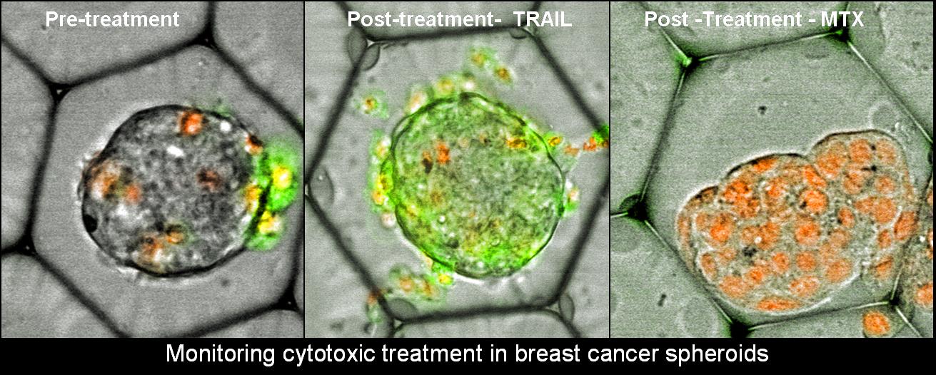
These cell models are used for the induction and detection of cell death (apoptosis) and cell proliferation.
Testing of biological compatibility and inertness
Different cell models are used to check the biological compatibility and inertness of the various CR prototypes, calibration and possible photo-damage of the present optical system.
Cell loading and rinsing
Cell loading procedures into the various CR types have been established for different cell type suspensions, at various concentrations. These include fluid manipulations in the CR preloaded with cells.
Cell maintaining and culturing
Individual cell suspensions (Jurkat T cells), as well as cells grown in mono-layers (PC3 cells) are cultured on different types of the Cell Retainer. We investigate the CR ability to preserve cells in their locations under routine medium exchanges and treatments with soluble agents.
Time-lapse viability studies are carried out, both with and without intermittent medium exchanges, employing both high and low cell densities on the CR. Such assessments include apoptosis, mitochondrial potential, membrane permeability, doubling time, mitotic index, and responsiveness to experimental stimuli (for example, intracellular Ca2+ response to T-cell receptor ligation).
Adherent cells (PC3) and individual cell suspensions (U937 cells) can be co-cultured on the same CR.
Â
Establishing diverse measurement capabilities on the CR
The following measurement capabilities are being developed utilizing the CR:
-
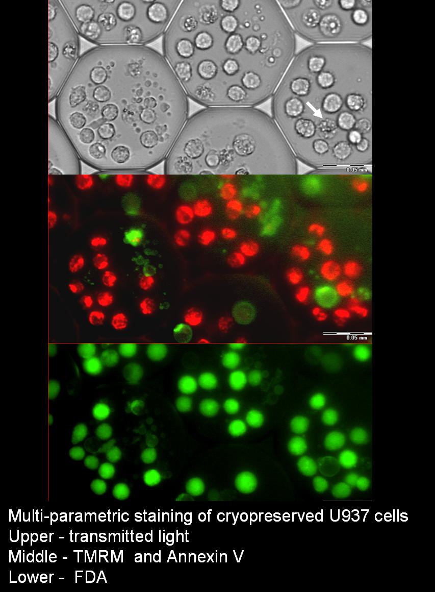
Surface marker staining of lymphocyte surface determinants (CDs multi-parametric measurements), Annexin V binding.
-
Intracellular protein staining (following fixation and permeabilization): intracellular Bcl2, Bax, various chemokines and cytokines.
-
Physiological assays multiplexed with antibody labeling assays. Cellular parameters include plasma membrane potential, mitochondrial potential, intracellular Ca2+, cAMP and ATP concentrations, enzyme activities, gene transcription, protein synthesis, cellular respiration, reactive oxygen species production, cell motility, cytokinesis. The labeling of intracellular proteins with fluorescent antibodies includes various chemokines and cytokines, pro- and anti-apoptotic protein expression. These measurement capabilities enable a direct correlation within the same cells, between vital functional parameters and features that require cell fixation for detection.
-
Cell staining and leakage (e.g. with FDA, RH123, DAF-2, DHR and DHE probes).
-
Chromatic staining.
-
Detection of rare cells.
-
Dynamic measurements:
-
Measurement of intracellular enzymatic reaction rates.
-
Determination of kinetic parameters (Km, Vmax).
-
Monitoring the staining kinetics of a mixed population of cells having different reaction rates (living and apoptotic cells, lymphocytes and monocytes).
-
Monitoring photo-oxidation reaction rates at a single cell level.
-
Photo-bleaching measurements.
-
Monitoring ROS and RNS generation rates at a single cell resolution.
-
Monitoring of intercellular interaction, visualization of T-cell mediated cytotoxicity.
-
Sub-population responses.
-