Medical R&D
- Cellular Endogenous Production of the Free Radical Species: Three-dimensional vs Two-dimensional Growth Mode
- Analysis of Cancer Cell Invasion and Anti-metastatic Drug Screening Using Hydrogel Micro-chamber Array (HMCA)-based Plates
- Discrimination of leukemic Jurkat cells from normal lymphocytes via novo label-free cytometry based on fluctuation of image gray values
- Measuring individual cell activation and invasion markers, to detect possible lymph node exposure to invasive breast cancer cells
- Information Theory based analytical models for cancer detection
- The involvement of Lysophosphatidylcholine (LPC) in lymphocyte apoptosis in atherosclerosis
- Differential immunosuppressive mechanisms of Methotrexate (MTX) and combined therapy in rheumatoid arthritis
- Improved classification of different pathological clones in bone marrow (BM) and peripheral blood (PB) of hemato-oncological patients
Cellular Endogenous Production of the Free Radical Species: Three-dimensional vs Two-dimensional Growth Mode
The complexity of tumor tissue, including cell-to-cell interaction, requires more accurate simulation of the natural tumor structure in vitro. Three-dimensional solid tumor models are more physiologically relevant and have a closer likeness to the native arrangement. This allows to overcome morphologic and functional alterations in the cell, due to non-physiological cultural 2D conditions in vitro. One of the most important advantages of the 3D multicellular objects' culturing is the more physiological response to microenvironmental signals including pharmaceutical treatment that can improve and accelerate the development and testing of a new drugs and therapeutic options.
This project is dedicated to better understand the nature of the most common and lethal primary brain tumor in adults - Glioblastoma Multiforme. The relationship between the level of endogenous NO and mitochondrial function in vitro at individual three-dimensional multicellular object resolution was studied. Previously, electrochemical measurements of NO production by glioblastoma cells in vitro revealed that A-172 cells, when cultured as traditional 2D monolayer, do not produce any detectable free radical species. Switching the growth mode to 3D conditions enables glioblastoma cells to produce endogenous NO in response to external stimulus, followed by mitochondrial hyperpolarization that can maintain the higher intracellular level of energy necessary for tumor cell activation, proliferation and movement, including cell invasion capacity, and thus, tumor metastasis. Moreover, considering the significant and widespread effects of NO in brain generally, and particularly in glioblastoma, modulation of NO levels is a feasible therapeutic strategy.
Analysis of Cancer Cell Invasion and Anti-metastatic Drug Screening Using Hydrogel Micro-chamber Array (HMCA)-based Plates
Cancer metastasis is known to cause 90% of cancer lethality. Metastasis is a multistage process which initiates with the penetration/invasion of tumor cells into neighboring tissue. Thus, invasion is a crucial step in metastasis, making the invasion process research and development of anti-metastatic drugs, highly significant. To address this demand, there is a need to develop 3D in vitro models which imitate the architecture of solid tumors and their microenvironment most closely to in vivo state on one hand, but at the same time be reproducible, robust and suitable for high yield and high content measurements. Currently, most invasion assays lean on sophisticated microfluidic technologies which are adequate for research but not for high volume drug screening. Other assays using plate-based devices with isolated individual spheroids in each well are material consuming and have low sample size per condition.
The goal of the current project is to provide a simple and reproducible biomimetic 3D cell-based system for the analysis of invasion capacity in large populations of tumor spheroids.
We developed a 3D model for invasion assay based on HMCA imaging plate for the research of tumor invasion and anti-metastatic drug discovery. This device enables the production of numerous uniform spheroids per well (high sample size per condition) surrounded by ECM components, while continuously and simultaneously observing and measuring the spheroids at single-element resolution for medium throughput screening of anti-metastatic drugs.
This platform was presented by the production of HeLa and MCF7 spheroids for exemplifying single cell and collective invasion. We compare the influence of the ECM component hyaluronic acid (HA) on the invasive capacity of collagen surrounding HeLa spheroids. Finally, we introduce Fisetin (invasion inhibitor) to HeLa spheroids and nitric oxide (NO) (invasion activator) to MCF7 spheroids. The results are analyzed by in-house software which enables semi-automatic, simple and fast analysis which facilitates multi-parameter examination.
Discrimination of leukemic Jurkat cells from normal lymphocytes via novo label-free cytometry based on fluctuation of image gray values
We introduce a simple, label-free cytometry technique, based on the spatio-temporal fluctuation analysis of pixel gray levels of a cell image utilizing the Gray Level Information Entropy (GLIE) function. In this study, the difference in GLIE random fluctuations and its biophysical etiology in a comparison cell model of leukemic Jurkat cells and human healthy donor lymphocytes was explored. A combination of common bright field microscopy and a unique imaging dish wherein cells are individually held untethered in a picoliter volume matrix of optical chambers was used. Random GLIE fluctuations were found to be greater in malignant Jurkat cells than in benign lymphocytes, while these fluctuations correlate with intracellular vesicle Mean Square Displacement (MSD) values and are inhibited by myosin-2 and adenosine triphosphate (ATP) inhibitors. These results suggest that the incoherent active forces acting on the cytoskeleton which cause mechanical dissipative fluctuation of the cytoskeletal and related intracellular content are the biophysical cellular mechanism behind the GLIE random fluctuation results. Analysis of the results in Jurkat cells and normal lymphocytes suggests the possible potential of this simple and automated label-free cytometry to identify malignancy, particularly in a diagnostic setup of multiple cell examination.
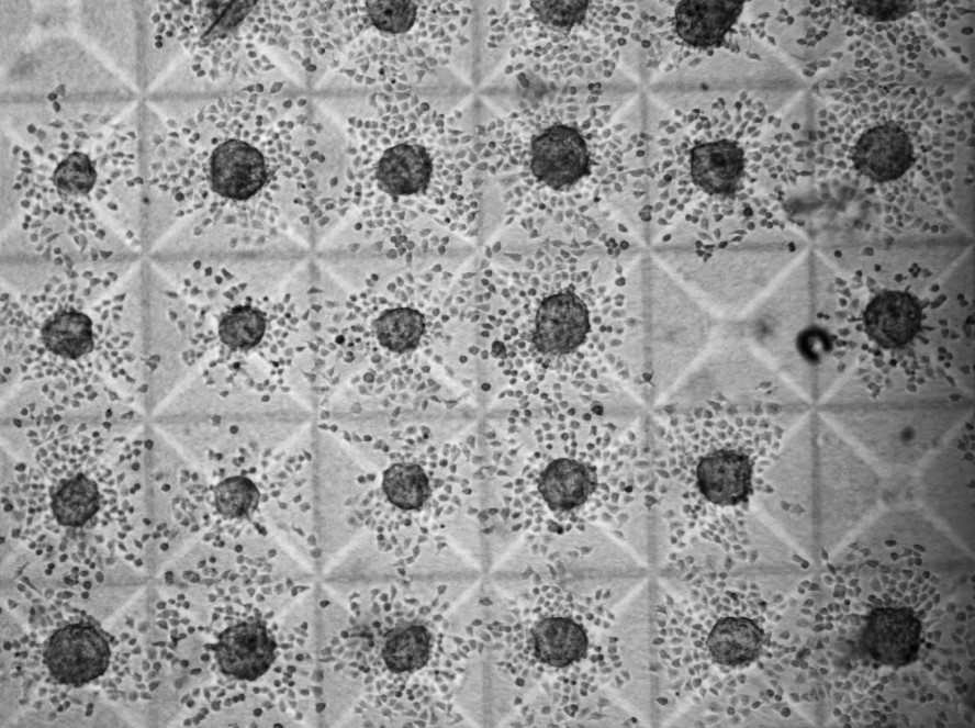
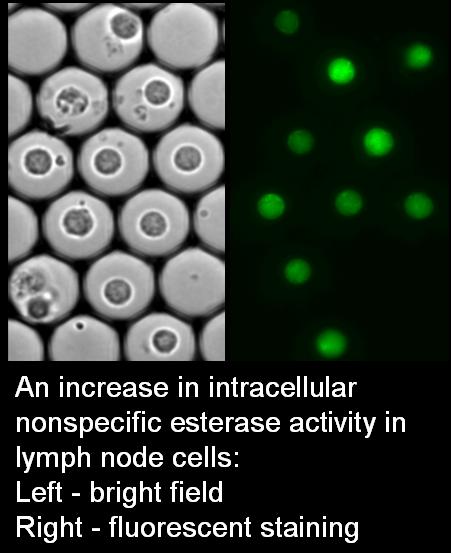
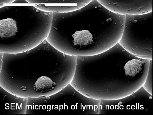
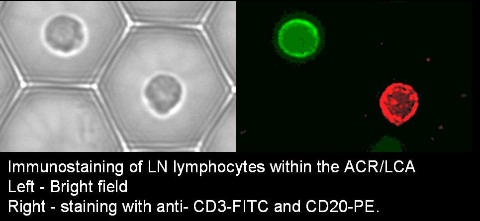
Measuring individual cell activation and invasion markers, to detect possible lymph node exposure to invasive breast cancer cells
Breast cancer often spreads from the primary tumor to regional lymph nodes. Lymph node (LN) status is one of the most important and widely used clinical predictors of cancer development, progression and survival rates. The aim of this study is to improve the sensitivity of the axillary lymph node (ALN) examination by adding new functional parameters to the standard pathological evaluation.
The Cell Retaining methodologies, as well as innovative systems enabling the collection and transfer of live LN cell samples from the hospital to the lab, are being used in this research project. High-content individual-cell assay protocols are being developed for intracellular reaction rates, the expression of the CD71 activation marker, and Matrix Metalloproteinases (MMPs) activity. The assays are being conducted on individual cells from tumor-free and metastatic LNs of cancer patients, under exposure to autologous tumor tissue. The analysis of live and post-fixation features of the same cell populations is revealing that the individual cells derived from metastatic lymph nodes are distinct from the cells derived from tumor-free nodes. The cell area, the rate of intracellular hydrolysis reaction and the membrane distribution of transferrin receptor, were found to be significantly different in cells from metastatic and non-metastatic nodes. These activation and invasion markers can be used to predict the LN status and to assess the involvement of LN in various cancerous processes (ovarian and lung cancer).
Information Theory based analytical models for cancer detection
This project concerns the construction of formal cellular diagnostic models for breast cancer detection, based on Information Theory that can be successfully applied in the research of heterogeneous biological systems, as it permits estimating non-linear relations between parameters.
The cellular diagnostic models employ individual living peripheral blood mononuclear cells (PBMC), derived from healthy subjects and cancer patients, under stimulation by the mitogen phytohemagglutinin (PHA) or by tumor tissue or under resting conditions. Using the Cell Retaining methodology, the individual cells can be fluorescently stained by fluorescein diacetate (FDA) and their fluorescence measured, at a single cell resolution, in successive time points, at different stain concentrations and at different wavelengths, generating diverse cell-based data patterns, including a variety of fluorescence polarization variants and individual cell Michaelis-Menten constants of FDA hydrolysis.
The measurement data are processed via Information Theory and non-parametric statistical analysis to determine the parameters and measurement conditions that can best discriminate between different groups. The normalized mutual information (uncertainty coefficient) is used as the measure of correlation/discrimination. An estimated general correlation has to be established between the parameters and the examined patterns (disease status) in the various bioassays. Ranking of the different groups of parameters permits to select the most informative (discriminative) parameters for cancer diagnosis.
When the Nearest neighbour rule is utilized for diagnosis, where the distance between elements is the weighted Hamming distance such formal cellular diagnostic models can concomitantly employ both quantitative input parameters (such as continuous fluorescence intensity values) and qualitative parameters (such as binary receptor markers). Also, decision trees and linear programming have been used for diagnosis, based on the boundaries established between data-sets. The diagnostic models that are being developed are open, i.e. they can accommodate new additional parameters, which may increase their effectiveness.
The involvement of Lysophosphatidylcholine (LPC) in lymphocyte apoptosis in atherosclerosis
LPC, an intermediate metabolite of phosphatidylcholine, is one of the major bioactive lipids, which has been identified in atherosclerotic plaque, and a major component of oxidized LDL (oxLDL), which plays a key role in atherosclerosis. T-lymphocytes found in early and late atherosclerotic lesions actively participate in lesion progression and rupture. The aim of this study is to evaluate the apoptotic effect of LPC on human lymphocytes. The CR methodology has been employed for multiple functional apoptotic assays followed by post-fixation studies. The functional cell-based assays include LPC stimulation of activated peripheral blood lymphocytes, as well as Jurkat T cells, and the analysis of apoptotic manifestations (production rates of reactive oxygen species - ROS, and intracellular Bax/Bcl-2 protein ratio). Real time kinetics of enhanced green fluorescent protein EGFP-Bid translocation has been measured under treatment of Jurkat-T cells transiently transfected with the pd4eGFP-Bid plasmid.
Additionally, preliminary results showed that LPC induces dose- and time-dependent changes in mitochondrial membrane potential, concurrently with an increase in ROS production rate, measured at an individual cell resolution. Moreover, peripheral lymphocytes derived from unstable and stable angina patients have been measured.
Differential immunosuppressive mechanisms of Methotrexate (MTX) and combined therapy in rheumatoid arthritis
The mechanism by which low dose methotrexate (the gold standard treatment for rheumatoid arthritis) or combined (MTX and anti-tumor necrosis factor) therapy exert their anti-inflammatory effects in rheumatoid arthritis (RA) patients is still debated. The aim of this project is to determine specific early events underlying the immunosuppressive mechanism of MTX therapy. Lately, the MTX immunosuppressive effect has been related to apoptosis, especially in active RA patients. Our preliminary data showed that MTX activity can be related to its induction of apoptosis with ROS involvement.
When focusing on MTX apoptotic mechanism, we use different cell based assays (including intracellular cytokines, cytokine receptors, ROS and NO assays, in relation to apoptosis), at an individual cell level using the CR methodologies. So far we showed that MTX induces IL-10 secretion, IL-4 and CCR4 receptor expression, and reduces CXCR3 expression in peripheral mononuclear cells derived from active RA patients, shifting the immune balance toward T-helper-2 (Th2) cell profile. The latter finding has a great relevance for RA patients who are characterized by Th1 predominance. The assessing of the MTX apoptotic mechanism, and its involvement with oxidative stress, in different subgroups of RA patients (active vs. non-active), may contribute to a better and more specific in vitro prediction of MTX efficiency.
Improved classification of different pathological clones in bone marrow (BM) and peripheral blood (PB) of hemato-oncological patients
There is sound evidence that genetic heterogeneity characterizes human neoplasias, despite the influence of their clonal origin and selection for tumor-specific genetic alterations. In the majority of human cancers, the pathological potential of genetic changes in small populations of cells is unknown. Apoptosis defects lead to the survival of the genetically abnormal cells and thus provide a setting for aggressive clone and tumor progress. The second negative aspect of the apoptotic mechanism is the appearance of multi-drug resistant cells that create clinical problems and impair the outcome of the patients treatment. The aim of the current study is to improve the classification of genetically different sub-clones of cancer cells by expanding the cytogenetic study via a correlation with functional live cell characteristics, at the level of individual cells. The utilization of the Cell Retaining technology will enable the correlation between chromosomal abnormalities and functional cellular characteristics in the same population of hematological cancer cells. Three models of cancer diseases expressing genetic heterogeneity are being investigated: chronic myelogenous leukemia (CML), multiple myeloma (MM) and pediatric acute lymphocytic leukemia (ALL). The ability to detect genetically different clones and to correlate with their functional apoptotic characteristics can provide a basis for prognosis and optimizing therapy for individual cancer patients. This can only be achieved by integrating cytogenetic analyses with the CR methodology.